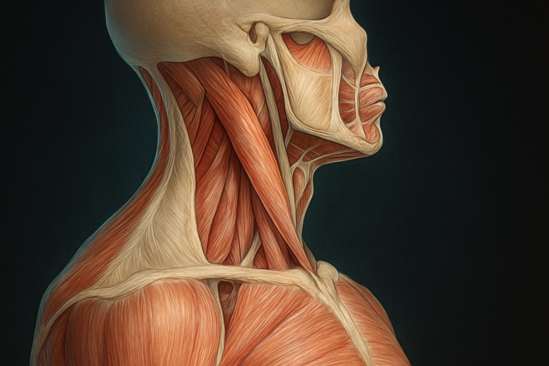
Spinal Stenosis Can Be Treated: Pathology and a Miracle Fascia Release Technique, from Zero
Original title (JP): 脊柱管狭窄症は治せる!病理学&奇跡の筋膜リリース手技テクニックをゼロから教えますね!
Supervised by
Shogo Fujii
Physiotherapist (PT) · Founder of the SHOGO FUJII® FASCIA METHOD
View profile →Clinical Article · 5 min read
Lumbar spinal stenosis, a common age-related condition, often leads to debilitating low back pain and leg symptoms. While traditional treatments frequently involve medication or surgery, many seek effective non-surgical alternatives. This article explores the pathology of spinal stenosis and introduces the promising role of fascial release for spinal stenosis, offering a unique perspective on managing its challenging symptoms.
Understanding Lumbar Spinal Stenosis: Causes and Symptoms
Spinal stenosis is the narrowing of the spinal canal, caused primarily by age-related degenerative changes like disc degeneration, facet joint hypertrophy, and thickening of the ligamentum flavum [1]. This reduces space for nerves, leading to compression and irritation. The hallmark symptom is neurogenic intermittent claudication – leg pain, heaviness, or numbness with walking/standing, relieved by sitting or leaning forward [2].
Spinal stenosis typically evolves gradually. While surgery is often an option, many patients don't experience lasting relief, highlighting the need for comprehensive, conservative approaches [3]. However, "red flag" symptoms like rapid muscle weakness or sudden bladder/bowel dysfunction necessitate immediate medical consultation.
The Pain Mechanism: More Than Just Compression
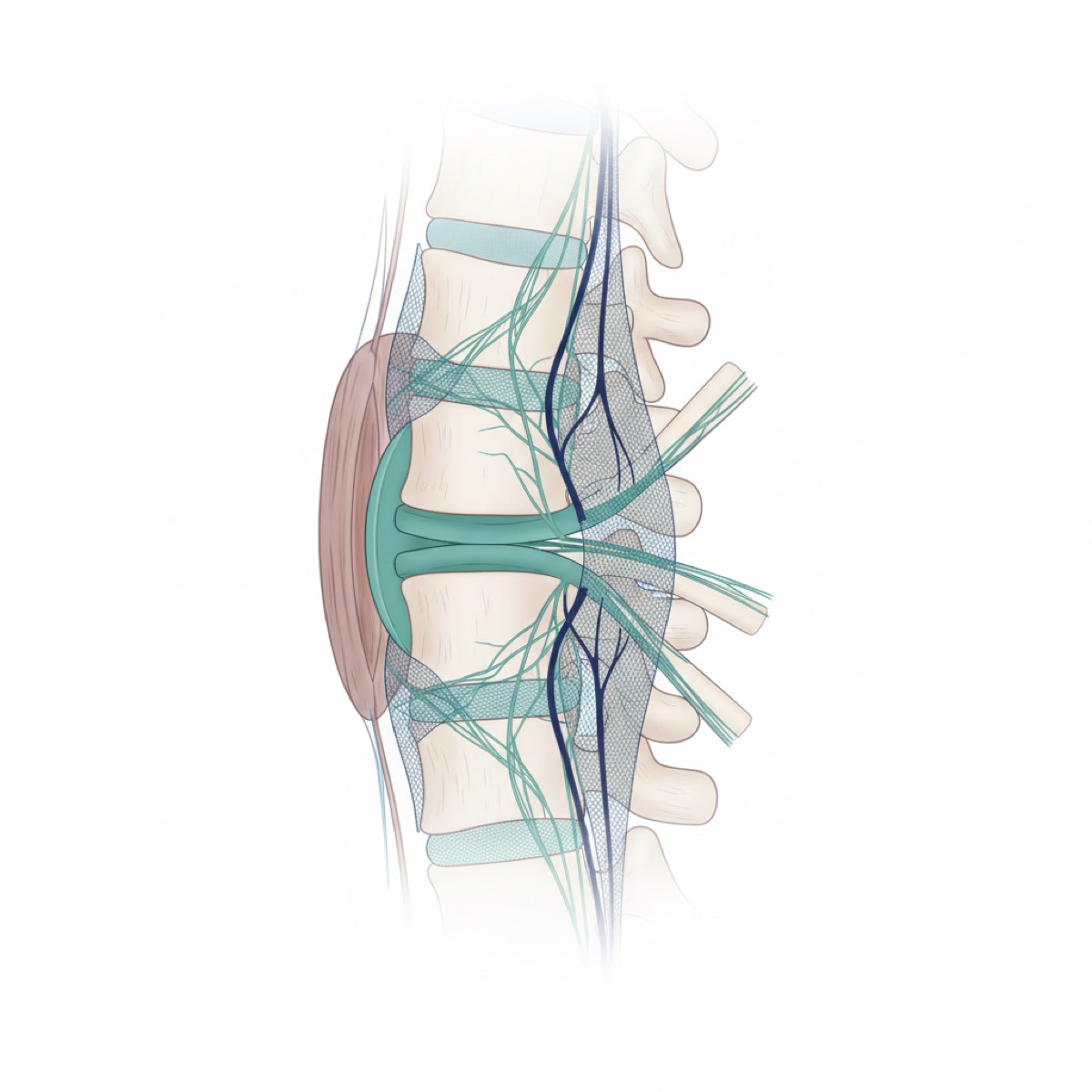
Pain and neurological symptoms stem from direct mechanical compression of nerves, reduced blood flow (ischemia), and local inflammation [4]. This combination irritates nerves, causing characteristic leg symptoms.
Posture profoundly influences symptoms. Lumbar extension (arching) narrows the canal, exacerbating pain. Flexion (leaning forward) often provides temporary relief by widening the canal and improving blood flow. This "flexion bias" is a key diagnostic indicator.
Navigating Diagnosis: The Nuance of Imaging and Clinical Assessment
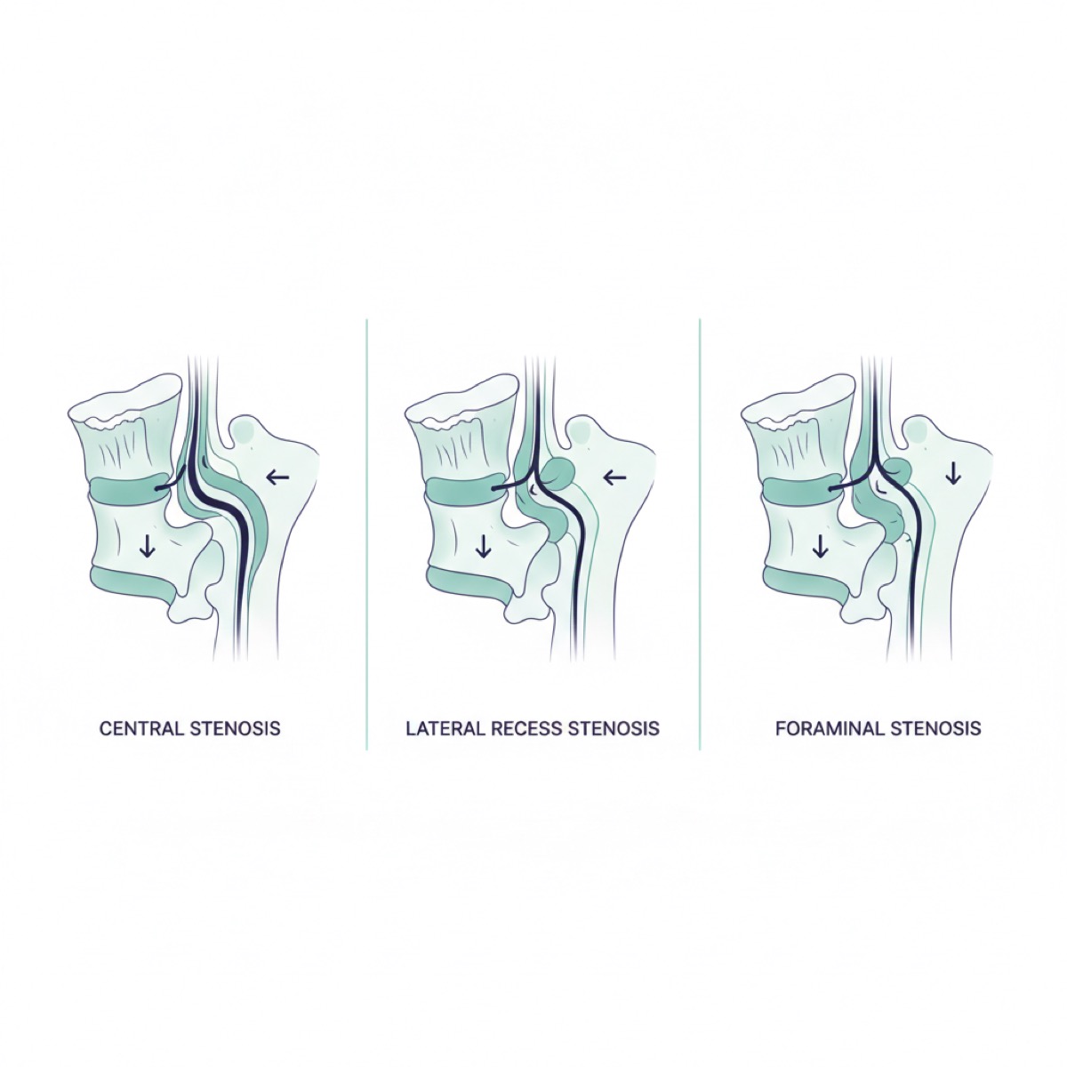
Spinal stenosis can manifest as central, lateral recess, or foraminal stenosis [5]. Imaging (X-rays, CT, MRI) reveals characteristic changes like ligamentum flavum hypertrophy, facet degeneration, and decreased disc height.
Critically, imaging findings don't always correlate with symptoms; stenosis degree also varies with body position. Therefore, a comprehensive clinical assessment of posture, movement, and functional limitations is paramount, complementing imaging results.
Fascial Release: A Non-Surgical Pathway for Spinal Stenosis Relief
Fascial release offers a promising non-surgical avenue. The philosophy acknowledges that while bony structure cannot be surgically altered via manual therapy, mechanical stress on nerves and soft tissues can be significantly reduced. The goal is to create a "flexion-biased" body posture, alleviating nerve compression and improving local circulation without invasive procedures.
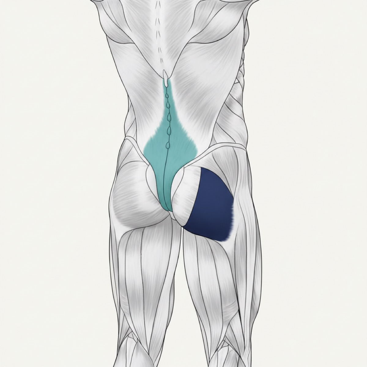
This strategy addresses anterior fascial lines and muscles that, when tight, contribute to excessive lumbar extension and increased spinal canal pressure. A key muscle is the psoas major, which, when shortened, pulls the lumbar spine into extension, exacerbating stenosis symptoms.
While direct psoas release is challenging, an effective indirect approach targets the tensor fasciae latae (TFL). The TFL, on the outer hip, originates from the anterior superior iliac spine (ASIS) and iliac crest. A unique fascial connection exists between the TFL and the psoas [6]. Applying precise manual pressure to the ASIS midpoint releases TFL tension, which in turn facilitates relaxation of the psoas and the anterior abdominal fascial complex. This significantly reduces mechanical forces contributing to spinal canal narrowing, improving leg numbness, intermittent claudication, and functional mobility.
Frequently asked questions
Q1 — What are the main symptoms of spinal stenosis? The primary symptom is neurogenic intermittent claudication: pain, numbness, or weakness in the legs that occurs with walking or standing and is relieved by sitting or leaning forward. Other symptoms may include low back pain, sciatica-like leg pain, and balance issues.
Q2 — Can spinal stenosis be treated without surgery? Yes, many cases can be effectively managed with conservative treatments like activity modification, pain medication, physical therapy, and manual therapies such as fascial release. Surgery is typically considered for severe cases, especially those with "red flag" symptoms like bladder/bowel dysfunction or progressive neurological deficits.
Q3 — How does fascial release help with spinal stenosis? Fascial release aims to reduce mechanical stress on spinal nerves and improve local blood flow by addressing tightness in muscles and fascia contributing to spinal compression. By releasing restricted anterior fascial lines, particularly through muscles like the psoas and indirectly via the tensor fasciae latae (TFL), these techniques help restore a more "flexion-biased" spinal posture, creating more space within the spinal canal and alleviating symptoms.
References
- [1]Ajimsha MS, Al-Mudahka NR, Al-Madzhar JA. Effectiveness of myofascial release: a systematic review of randomized controlled trials. J Bodyw Mov Ther. 2015;19(1):102–112.
- [2]Tozzi P, Bongiorno D, Vitturini C. Fascial release effects on patients with non-specific cervical or lumbar pain. J Bodyw Mov Ther. 2011;15(4):405–416.
- [3]Zügel M, Maganaris CN, Wilke J, et al. Fascial tissue research in sports medicine: from molecules to tissue adaptation, injury and diagnostics: consensus statement. Br J Sports Med. 2018;52(23):1497.
- [4]Klingler W, Velders M, Hoppe K, Pedro M, Schleip R. Clinical relevance of fascial tissue and dysfunctions. Curr Pain Headache Rep. 2014;18(8):439.
- [5]Wilke J, Vogt L, Niederer D, Banzer W. Is remote stretching based on myofascial chains as effective as local exercise? A randomised-controlled trial. J Sports Sci. 2017;35(20):2021–2027.
- [6]Palomeque-del-Cerro L, Arráez-Aybar LA, Rodríguez-Blanco C, et al. A systematic review of the soft-tissue connections between neck muscles and dura mater: the myodural bridge. Spine. 2017;42(1):49–54.
Related videos
Palpation X-Ray #84 | Is the Sacrum Really a Flat Plate? It's a Wedge, Wide at the Top and Narrowing Downward — Trace Down from the Lumbar Spine and Confirm at Gluteus Maximus
0:56
Dramatic Improvement in One Session: Manual Approach to the Pelvic Girdle, with Palpation of Anatomical Landmarks and Movement Assessment Before and After (Hands-On Demo)
5:48