THE METHOD

The science behind the method.

Fascia-based manual therapy reads the body as one connected system rather than a collection of separate parts. The framework below sets out the reasoning behind the Fujii Method and the peer-reviewed science that informs each step. It is an educational overview for clinicians and is not medical advice; it describes mechanisms and evidence, not guaranteed outcomes.

21 peer-reviewed references/Evidence-informed/Quantified with elastography

FOUNDATION

Fascia is one continuous, sensing network
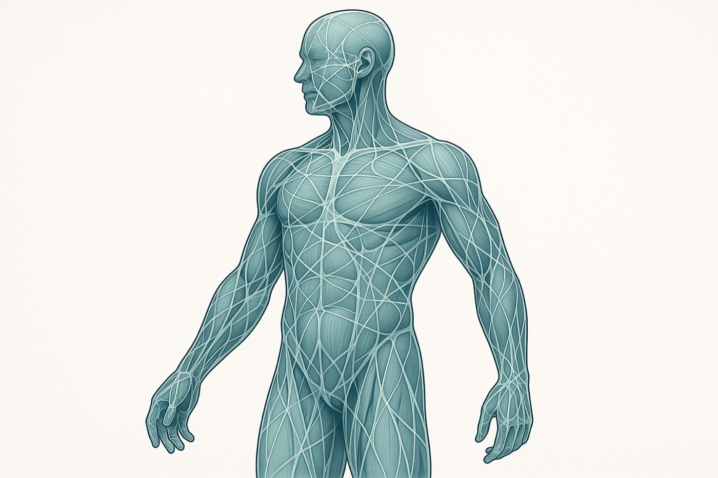
Diagram of the fascia connective-tissue network wrapping and linking muscles throughout the body

Fascia is the connective-tissue network that wraps and links every muscle, organ and joint. It is increasingly understood not as inert packing material but as a body-wide, mechanically continuous and richly innervated signalling system [1, 2]. This continuity is why a restriction in one region can change load and sensation in another.

At the tissue level, the gliding behaviour of fascia depends on its hydration and the composition of the loose connective tissue between layers — including hyaluronan, whose altered state has been implicated in the genesis of myofascial pain [3]. A 2018 international consensus statement in the British Journal of Sports Medicine concluded that fascial tissue is an adaptable, clinically relevant structure that responds to mechanical load [4].
PRINCIPLE 1

Myofascial chains: the source is rarely the symptom

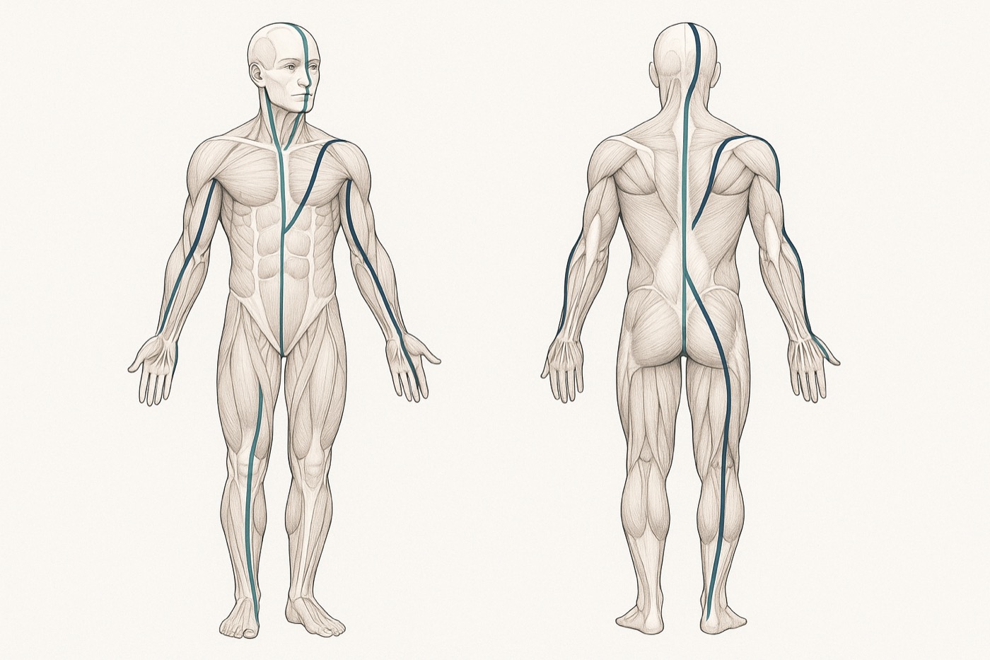
Diagram of myofascial chains (anatomy lines) running as continuous lines of pull across the body

Muscles do not act in isolation. Force is transmitted along myofascial chains — the so-called "anatomy lines" — so tension generated in one segment can be carried mechanically to distant regions [5, 6]. A systematic review of these chains found consistent anatomical evidence for several continuous lines of pull across the body [5].

This connectivity is not only anatomical but functional: a randomised controlled trial showed that stretching the lower limb increased range of motion in the cervical spine, consistent with force transmission along a myofascial chain [7]. Clinically, this is why the Fujii Method assesses the whole chain and treats the true source of restriction rather than only the site where pain is felt.

PRINCIPLE 2

The central axis: cranium, spine and pelvis as one line

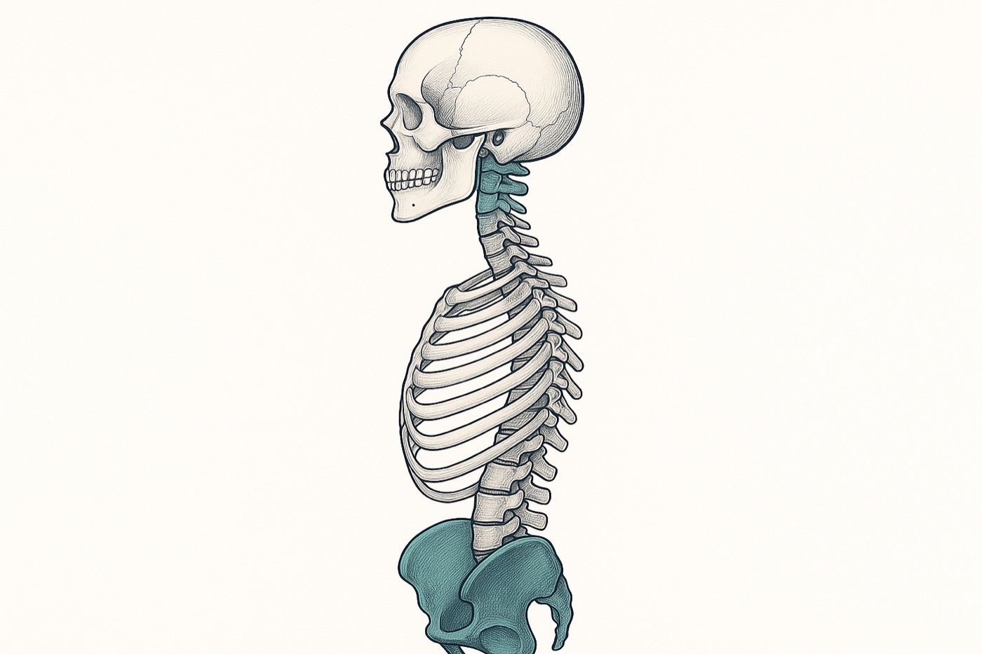
Diagram of the central body axis aligning the cranium, cervical spine, spine and pelvis

The second principle works with the body's central axis — the line that runs from the occiput and upper cervical spine through the spine to the pelvis. There is a genuine anatomical basis for treating the craniocervical junction as continuous with deeper structures: a systematic review documented direct soft-tissue connections (the "myodural bridge") between the suboccipital muscles and the dura mater [8].

Because fascia is mechanically continuous [2], restoring alignment and glide along this axis can influence tension far from the point of contact. The Fujii Method restores the axis first — pelvis, spine and craniocervical junction — then re-tests how distal symptoms respond.

EVIDENCE

Releasing fascia by hand: what controlled trials show

Hands-on fascial techniques have been tested in controlled research. A systematic review of randomised controlled trials concluded that myofascial release is a promising intervention for a range of conditions, while calling for larger, higher-quality studies [9].

For spinal pain specifically, a randomised, sham-controlled trial reported that fascial release reduced pain and improved function in patients with non-specific cervical or lumbar pain [10]. Evidence-informed practice means treating these results as support for a careful, reasoned approach — not as a promise of a fixed outcome.

PRINCIPLE 3

Persistent pain lives partly in the nervous system

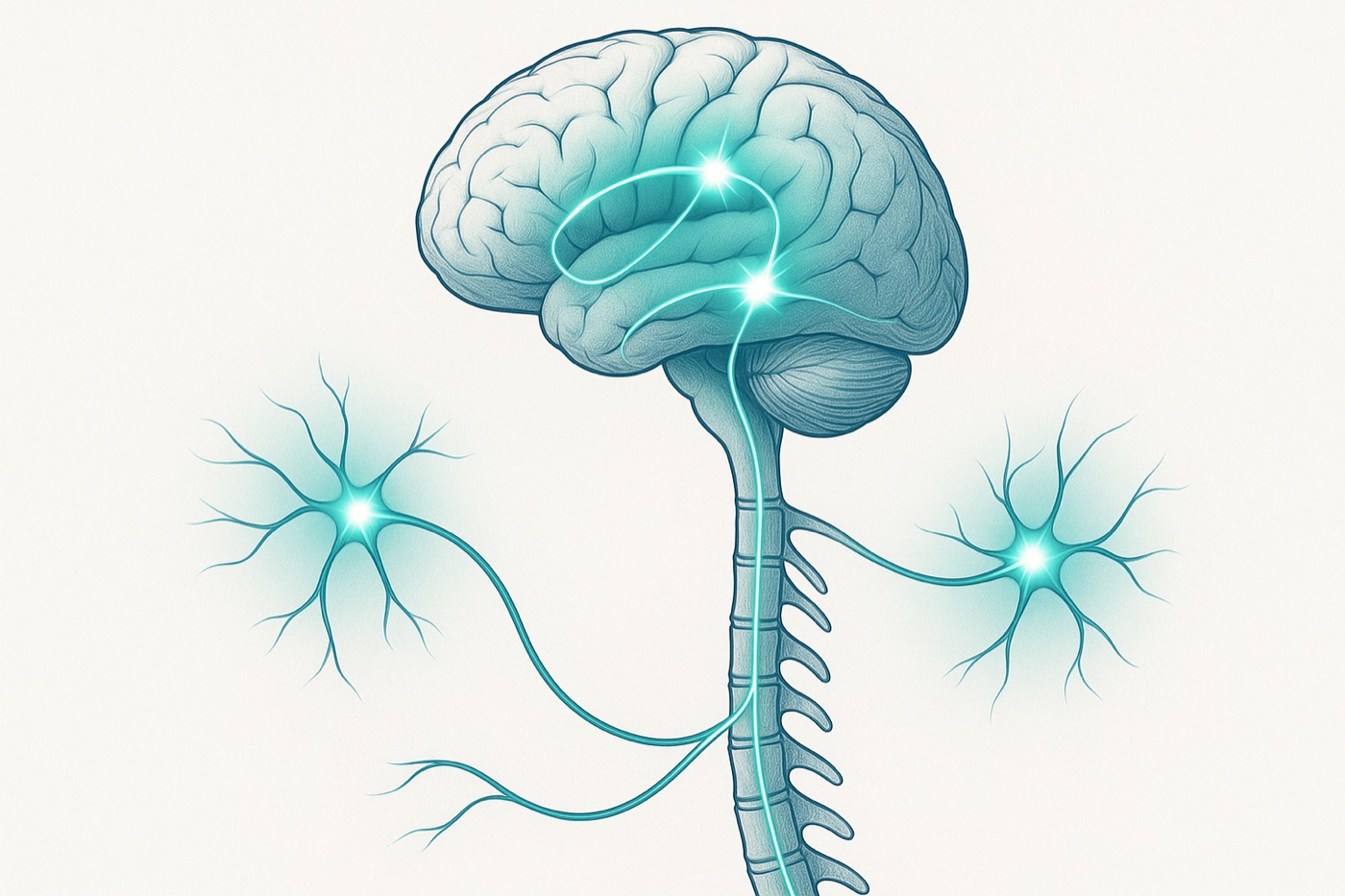
Diagram of central sensitization: amplified pain signalling in the spinal cord and brain

When pain becomes chronic, the nervous system itself changes. Central sensitization — an amplification of signalling in the central nervous system — can make pain persist even after the original tissue issue has settled [11]. This is why pain that is only chased at the painful spot, with heat or massage, can keep returning.

A method that accounts for the nervous system, rather than tissue alone, is therefore better matched to how persistent pain actually behaves [11]. This reasoning underpins the Fujii Method's attention to the whole system, not just the symptomatic muscle.

PRINCIPLE 3

The autonomic shift: from guarding to recovery

A guarded, sympathetically driven state keeps muscles braced and tissue sensitised. The parasympathetic (vagal) system does the opposite — and it is also a brake on inflammation. The "inflammatory reflex" describes how vagal activity down-regulates pro-inflammatory signalling [12], and stimulating this pathway has been shown to reduce cytokine production and disease activity in a human trial in rheumatoid arthritis [13].

Calm, deliberate manual input and breathing are used to encourage a shift toward this recovery state, so that deeper tissue can release. This is presented as a physiological rationale, not a claim to treat any specific disease.

PRINCIPLE 3

The gut–brain axis and pain modulation

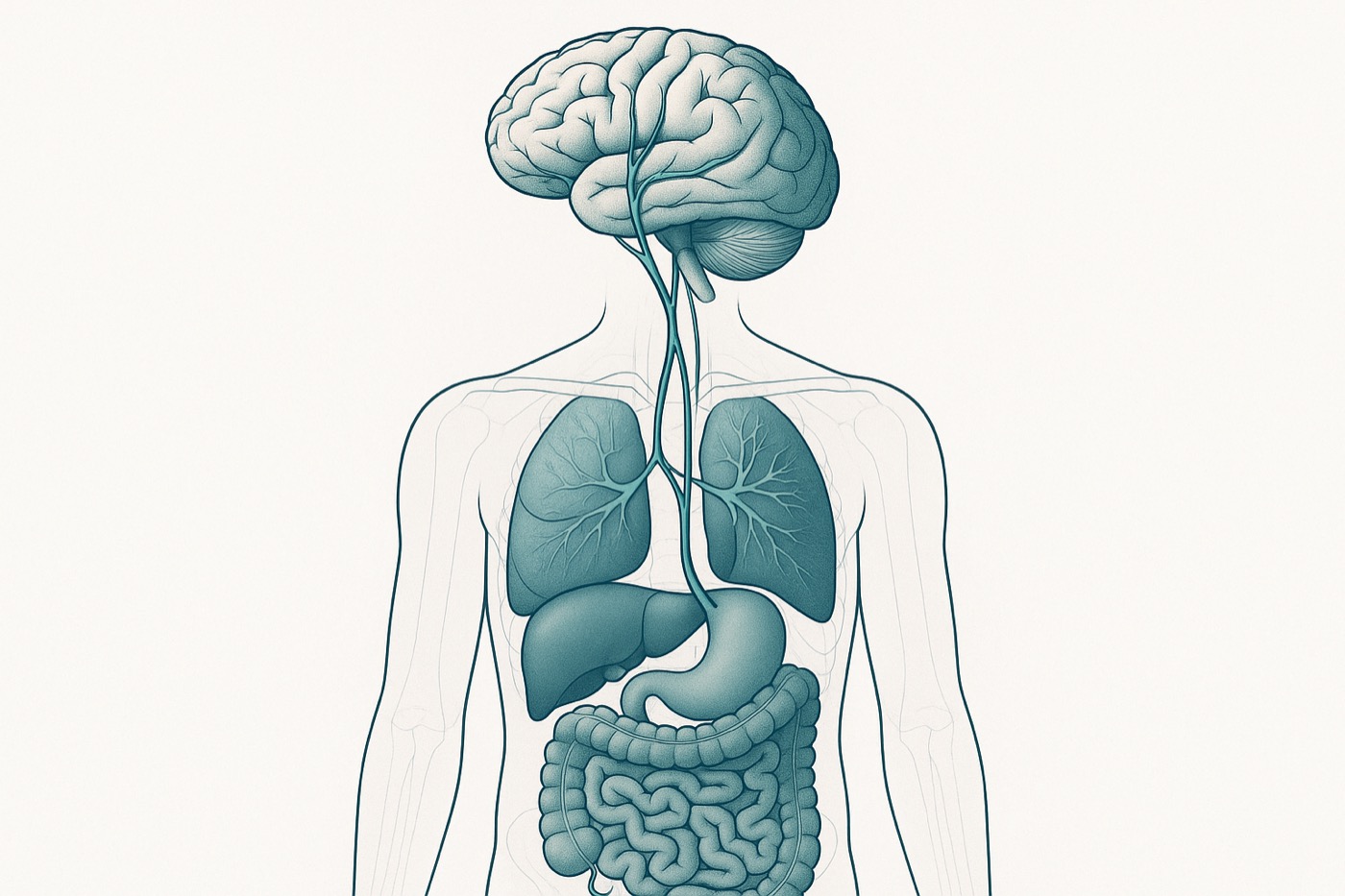
Diagram of the gut–brain axis: the vagus nerve linking the brain, viscera and gut

The gut and brain are in constant two-way communication along the gut–brain axis [14], and the gut microbiota influence this signalling and behaviour [15]. A growing body of work links the gut microbiome to the regulation of pain, describing plausible molecular pathways and therapeutic potential [16].

This is why the Fujii Method considers the viscera and gut state as part of the picture in persistent cases. The science here is emerging rather than settled, and it is offered as a rationale for a whole-person approach — not as a treatment claim.

SUPPORT

Pain chemistry, hydration and clearance

Pain and inflammation are partly chemical. Prostaglandins are key lipid mediators that sensitise nerve endings and drive inflammatory pain [17]. Supporting the body's normal circulation and clearance — including adequate hydration — is a sensible adjunct to manual work, given fascia's dependence on tissue fluid for healthy glide [3].

General hydration guidance should always be matched to the individual and any medical conditions; it complements, and never replaces, appropriate medical care.

SUPPORT

Nutrition as an anti-inflammatory lever

Diet can modulate the inflammatory load that maintains pain. A systematic review with meta-analysis found that dietary interventions can reduce pain in people with chronic pain [20]. Among plant compounds, the flavonoid apigenin — abundant in celery and parsley — has well-documented anti-inflammatory and analgesic activity in preclinical research [18], and celery (Apium graveolens) has a body of work describing its antioxidant and anti-inflammatory properties [19].

These are presented as evidence-informed nutritional supports, not as drug substitutes. Any change for a specific condition should be discussed with a qualified professional.

RIGOR

Measured, not guessed: imaging the change

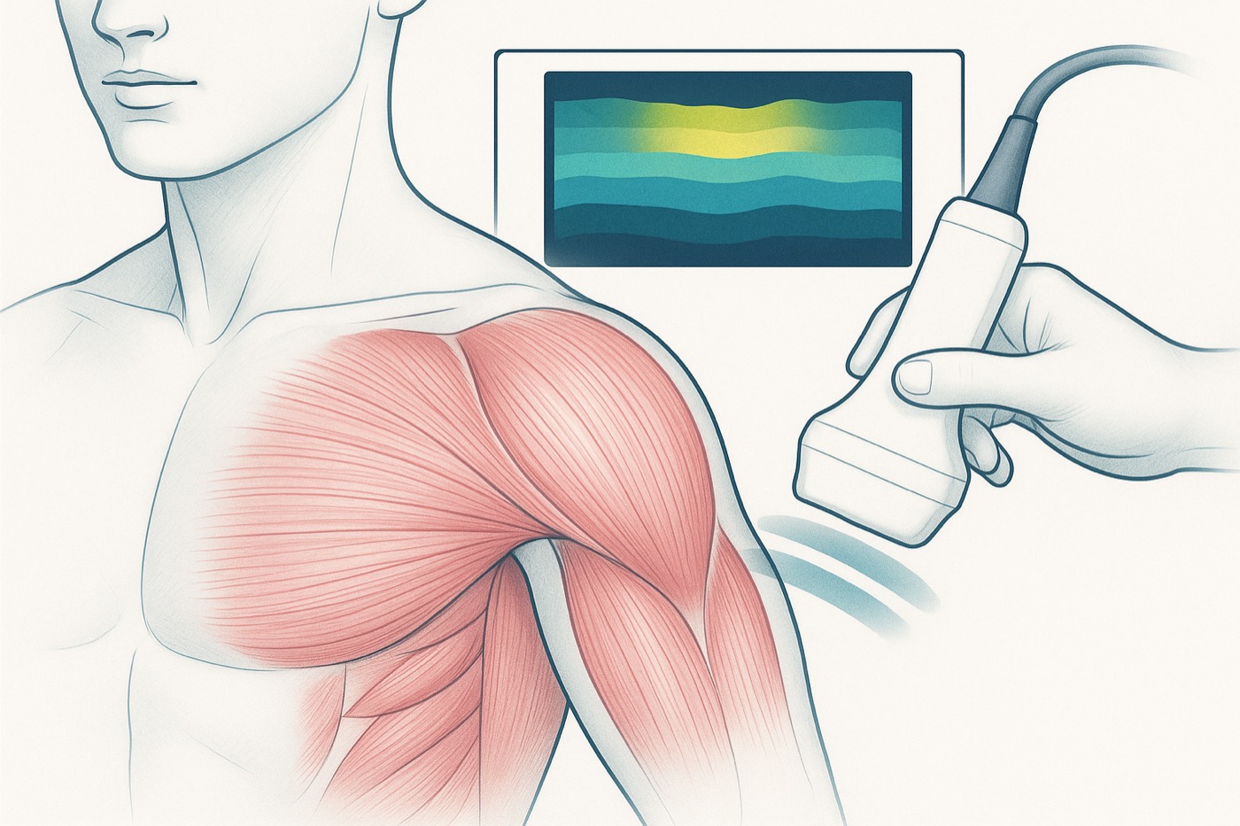
Diagram of ultrasound shear-wave elastography mapping soft-tissue stiffness over muscle

What can be measured can be improved. Ultrasound shear-wave elastography now allows clinicians to quantify tissue stiffness objectively, with established musculoskeletal applications [21]. Shogo Fujii's own research applies elastography to quantify changes in muscular rigidity after manual therapy — moving outcomes from subjective impression toward measurable change.

This commitment to measurement is what separates a reasoned method from technique performed on faith.

FREQUENTLY ASKED QUESTIONS

What is fascia-based manual therapy?

Fascia is the continuous connective-tissue network that wraps and links every muscle, organ and joint. Fascia-based manual therapy works with this network — locating densified, restricted areas and treating them by hand — rather than addressing only the spot where pain is felt.

What makes this approach different?

Instead of chasing symptoms, the Fujii Method reasons about how restrictions transmit tension across the fascial chains (the “anatomy lines”). Treatment is selected from that map, and increasingly verified with imaging such as ultrasound elastography.

Why a hands-on, non-pharmacological approach?

Many movement-related complaints stem from how soft tissue glides and loads, not from a single structure. A skilled, drug-free manual approach can address those mechanics directly — as an evidence-informed complement to medical care, never a replacement for it.

How is a treatment reasoned out?

Assess the movement and the fascial chains involved, find the key restricted points, treat by hand, then re-test the movement. The videos on this site break down that assessment-treat-retest loop technique by technique.

Who is this for?

Physiotherapists, manual therapists, bodyworkers and clinicians who want to deepen their hands-on skills. The content is educational and intended to support qualified practice — it is not medical advice for self-treatment.

REFERENCES

  1. 1.Langevin HM. Connective tissue: a body-wide signaling network? Med Hypotheses. 2006;66(6):1074–1077. doi:10.1016/j.mehy.2005.12.032
  2. 2.Schleip R. Fascial plasticity — a new neurobiological explanation. J Bodyw Mov Ther. 2003;7(1):11–19. doi:10.1016/S1360-8592(02)00067-0
  3. 3.Stecco C, Stern R, Porzionato A, et al. Hyaluronan within fascia in the etiology of myofascial pain. Surg Radiol Anat. 2011;33(10):891–896. doi:10.1007/s00276-011-0876-9
  4. 4.Zügel M, Maganaris CN, Wilke J, et al. Fascial tissue research in sports medicine: consensus statement. Br J Sports Med. 2018;52(23):1497. doi:10.1136/bjsports-2018-099308
  5. 5.Wilke J, Krause F, Vogt L, Banzer W. What is evidence-based about myofascial chains? A systematic review. Arch Phys Med Rehabil. 2016;97(3):454–461. doi:10.1016/j.apmr.2015.07.023
  6. 6.Krause F, Wilke J, Vogt L, Banzer W. Intermuscular force transmission along myofascial chains: a systematic review. J Anat. 2016;228(6):910–918. doi:10.1111/joa.12464
  7. 7.Wilke J, Vogt L, Niederer D, Banzer W. Is remote stretching based on myofascial chains as effective as local exercise? A randomised-controlled trial. J Sports Sci. 2017;35(20):2021–2027. doi:10.1080/02640414.2016.1251606
  8. 8.Palomeque-del-Cerro L, Arráez-Aybar LA, Rodríguez-Blanco C, et al. A systematic review of the soft-tissue connections between neck muscles and dura mater: the myodural bridge. Spine. 2017;42(1):49–54. doi:10.1097/BRS.0000000000001907
  9. 9.Ajimsha MS, Al-Mudahka NR, Al-Madzhar JA. Effectiveness of myofascial release: a systematic review of randomized controlled trials. J Bodyw Mov Ther. 2015;19(1):102–112. doi:10.1016/j.jbmt.2014.06.001
  10. 10.Tozzi P, Bongiorno D, Vitturini C. Fascial release effects on patients with non-specific cervical or lumbar pain. J Bodyw Mov Ther. 2011;15(4):405–416. doi:10.1016/j.jbmt.2010.11.003
  11. 11.Woolf CJ. Central sensitization: implications for the diagnosis and treatment of pain. Pain. 2011;152(3 Suppl):S2–S15. doi:10.1016/j.pain.2010.09.030
  12. 12.Tracey KJ. The inflammatory reflex. Nature. 2002;420(6917):853–859. doi:10.1038/nature01321
  13. 13.Koopman FA, Chavan SS, Miljko S, et al. Vagus nerve stimulation inhibits cytokine production and attenuates disease severity in rheumatoid arthritis. Proc Natl Acad Sci USA. 2016;113(29):8284–8289. doi:10.1073/pnas.1605635113
  14. 14.Mayer EA. Gut feelings: the emerging biology of gut–brain communication. Nat Rev Neurosci. 2011;12(8):453–466. doi:10.1038/nrn3071
  15. 15.Cryan JF, Dinan TG. Mind-altering microorganisms: the impact of the gut microbiota on brain and behaviour. Nat Rev Neurosci. 2012;13(10):701–712. doi:10.1038/nrn3346
  16. 16.Guo R, Chen LH, Xing C, Liu T. Pain regulation by gut microbiota: molecular mechanisms and therapeutic potential. Br J Anaesth. 2019;123(5):637–654. doi:10.1016/j.bja.2019.07.026
  17. 17.Ricciotti E, FitzGerald GA. Prostaglandins and inflammation. Arterioscler Thromb Vasc Biol. 2011;31(5):986–1000. doi:10.1161/ATVBAHA.110.207449
  18. 18.Salehi B, Venditti A, Sharifi-Rad M, et al. The therapeutic potential of apigenin. Int J Mol Sci. 2019;20(6):1305. doi:10.3390/ijms20061305
  19. 19.Kooti W, Daraei N. A review of the antioxidant activity of celery (Apium graveolens L). J Evid Based Complementary Altern Med. 2017;22(4):1029–1034. doi:10.1177/2156587217717415
  20. 20.Field R, Pourkazemi F, Turton J, Rooney K. Dietary interventions are beneficial for patients with chronic pain: a systematic review with meta-analysis. Pain Med. 2021;22(3):694–714. doi:10.1093/pm/pnaa378
  21. 21.Taljanovic MS, Gimber LH, Becker GW, et al. Shear-wave elastography: basic physics and musculoskeletal applications. RadioGraphics. 2017;37(3):855–870. doi:10.1148/rg.2017160116

This page is an educational overview of mechanisms and evidence. It is not medical advice and makes no guarantee of any individual outcome. Nutrition and hydration guidance is general and should be adapted with a qualified professional; manual therapy is a complement to, not a replacement for, appropriate medical care.

FREE DOWNLOAD

Free Guide: The Anatomy-Line Fascia Release Starter Kit

A step-by-step introduction to the fascia-release techniques Shogo Fujii teaches to clinicians worldwide. Enter your email to get it instantly.

By signing up you agree to our Privacy Policy. You can unsubscribe at any time.