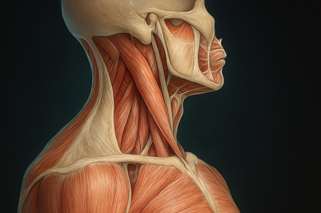
Palpation X-Ray #1: Why 90% Never Actually Find the Psoas Major — Navel-ASIS Midpoint, Aortic Pulse, and Differentiating from Iliacus
Original title (JP): 触診X線 #1|大腰筋を触れてない9割の理由|臍とASISの中点・大動脈の拍動・腸骨筋との鑑別 #藤井翔悟 #Shorts

Supervised by
Shogo Fujii
Physiotherapist (PT) · Founder of the SHOGO FUJII® FASCIA METHOD
View profile →Clinical Article · 5 min read
Psoas major palpation is one of the most misunderstood skills in manual therapy, because the muscle most clinicians press on the abdomen is actually the iliacus, not the psoas. The true psoas major sits deeper, running from the sides of the T12–L5 vertebrae down toward the lesser trochanter of the femur. Getting the landmark, the depth, and the safety checks right changes what you're actually assessing and treating. This guide walks through where to place your fingers, how to avoid the abdominal aorta, and how to confirm you're on the correct muscle before drawing any clinical conclusions.[1]
Why Is Psoas Major Palpation So Hard to Get Right?
Under the skin of the lateral abdomen, most therapists press directly onto a firm band that they assume is the psoas. In reality, that superficial band is usually the iliacus, a separate muscle that only merges with the psoas near its insertion to form the iliopsoas complex.[2]
The psoas major itself originates from the transverse processes and vertebral bodies of T12 through L5, then travels down through the pelvis to insert near the lesser trochanter of the femur. Because it lies deep to the abdominal wall and bowel, it cannot be palpated the same way you'd palpate a superficial muscle — depth, angle, and landmark all matter.[3]
Where Exactly Do You Find the Psoas Muscle?
Start at the midpoint between the navel and the ASIS (anterior superior iliac spine). This point sits just lateral to the border of the rectus abdominis and serves as your entry path from the front of the abdomen toward the vertebral column.
With the hip and knee slightly flexed to slacken the abdominal wall, let your fingers sink slowly toward the side of the lumbar vertebral bodies rather than pressing straight down. Go slowly — sudden deep pressure here is uncomfortable and unnecessary.
How Do You Confirm You're on the Psoas, Not the Iliacus?
Before going any deeper, pay attention to pulsation. If you feel a strong pulse, especially on the left side, that is the abdominal aorta — shift your fingers slightly lateral before continuing. This is a basic safety check that should never be skipped during psoas major palpation.[4]
Once positioned safely, ask the person to flex the hip against light resistance. If the tissue under your fingers tenses at this landmark near the spine, you are likely on the psoas. If the sensation of tightening shows up instead in the groin or inguinal region, you're feeling the iliacus rather than the psoas.[5]
What Does Fascia Have to Do with Psoas Tightness?
Interestingly, direct pressure on the psoas isn't always the preferred approach. The psoas major connects functionally to the adductor longus of the inner thigh through a fascial pathway often described as part of the deep front line.[6]
Because of this connection, working on the adductor longus can produce a measurable change in psoas tension without ever touching the abdomen directly. A simple way to check this is the Thomas test, performed before and after addressing the adductor longus, comparing hip extension range on each side.
Frequently asked questions
Is psoas major palpation safe to try on yourself? — Light, slow exploration at the described landmark is generally low-risk for most people, but anyone with abdominal pain, a pulsatile mass, pregnancy, or a known aortic condition should avoid deep abdominal palpation and consult a qualified clinician instead; this article is educational and not medical advice.
Why does the psoas matter for low back pain? — Because the psoas major attaches directly onto the lumbar vertebral bodies and crosses the hip joint, chronic tightness or poor control in this muscle is frequently discussed as a contributing factor in low back and hip discomfort, though it is rarely the sole cause and should be assessed alongside other structures.[3]
Can working on a different muscle really change psoas tension? — Yes, this is consistent with fascial-chain concepts like the deep front line, where tension can transmit between connected muscles such as the adductor longus and the psoas; results vary between individuals and are best confirmed with a simple before-and-after test like the Thomas test rather than assumed.
References
- [1]Ajimsha MS, Al-Mudahka NR, Al-Madzhar JA. Effectiveness of myofascial release: a systematic review of randomized controlled trials. J Bodyw Mov Ther. 2015;19(1):102–112.
- [2]Tozzi P, Bongiorno D, Vitturini C. Fascial release effects on patients with non-specific cervical or lumbar pain. J Bodyw Mov Ther. 2011;15(4):405–416.
- [3]Zügel M, Maganaris CN, Wilke J, et al. Fascial tissue research in sports medicine: from molecules to tissue adaptation, injury and diagnostics: consensus statement. Br J Sports Med. 2018;52(23):1497.
- [4]Klingler W, Velders M, Hoppe K, Pedro M, Schleip R. Clinical relevance of fascial tissue and dysfunctions. Curr Pain Headache Rep. 2014;18(8):439.
- [5]Wilke J, Vogt L, Niederer D, Banzer W. Is remote stretching based on myofascial chains as effective as local exercise? A randomised-controlled trial. J Sports Sci. 2017;35(20):2021–2027.
- [6]Palomeque-del-Cerro L, Arráez-Aybar LA, Rodríguez-Blanco C, et al. A systematic review of the soft-tissue connections between neck muscles and dura mater: the myodural bridge. Spine. 2017;42(1):49–54.
Related videos
Palpation X-Ray #84 | Is the Sacrum Really a Flat Plate? It's a Wedge, Wide at the Top and Narrowing Downward — Trace Down from the Lumbar Spine and Confirm at Gluteus Maximus
0:56
Dramatic Improvement in One Session: Manual Approach to the Pelvic Girdle, with Palpation of Anatomical Landmarks and Movement Assessment Before and After (Hands-On Demo)
5:48