Keep This: Gluteus Minimus Release, the Hidden Key to Better Lower Back Pain
Original title (JP): 【保存版】小臀筋リリース――腰痛改善の“隠れキャラ”を暴くテクニック

Supervised by
Shogo Fujii
Physiotherapist (PT) · Founder of the SHOGO FUJII® FASCIA METHOD
View profile →Clinical Article · 6 min read
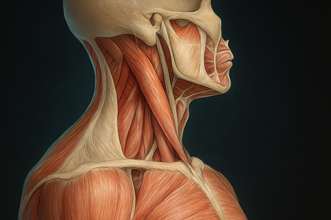
Persistent low back pain can often stem from unexpected sources, and one such area frequently overlooked is the gluteus minimus muscle. This deep-seated gluteal muscle plays a crucial, yet often underestimated, role in hip stability and can contribute significantly to symptoms of **gluteus minimus low back pain relief**. Understanding its function and how to effectively address its dysfunction through techniques like myofascial release is key for both clinicians and informed patients seeking lasting relief [1].
Why Your Gluteus Minimus Might Be Causing Your Low Back Pain
The gluteus minimus, though smaller than its gluteal counterparts, is a powerful hip abductor and internal rotator, vital for stabilizing the pelvis during walking and standing. When this muscle becomes tight, dysfunctional, or harbors trigger points, it can refer pain patterns that mimic or exacerbate low back pain, often extending into the hip, buttock, and even down the leg [2]. Its deep location means its involvement is sometimes missed in standard assessments, making targeted intervention crucial for effective **myofascial release for low back pain**.
Dysfunction in the gluteus minimus can compromise the intricate biomechanics of the lumbopelvic region, leading to compensatory movements and increased stress on the lower back. Addressing this often-overlooked muscle is a key strategy in comprehensive **hip-related low back pain** management, offering a pathway to improved function and reduced discomfort [3].
The Crucial Role of Accurate Palpation in Myofascial Release
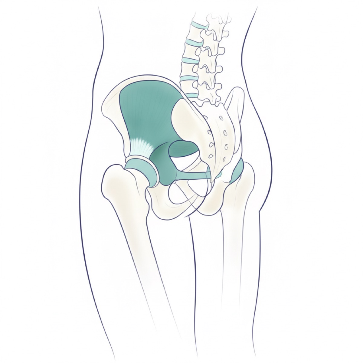
Effective myofascial release hinges on the ability to precisely locate and differentiate the target muscle tissue. For a deep muscle like the gluteus minimus, accurate palpation is not just important—it's foundational. Without a clear understanding of its anatomical position and surrounding structures, attempts at treatment may be misdirected, leading to ineffective outcomes or even discomfort in unintended areas [4].
Clinicians must develop a keen sense of touch and a solid anatomical knowledge base to confidently identify the gluteus minimus. This skill ensures that the therapeutic pressure is applied directly to the dysfunctional tissue, maximizing the potential for releasing tension and restoring normal muscle function, which is paramount for achieving **gluteus minimus low back pain relief**.
Mastering Gluteus Minimus Palpation: Key Anatomical Landmarks
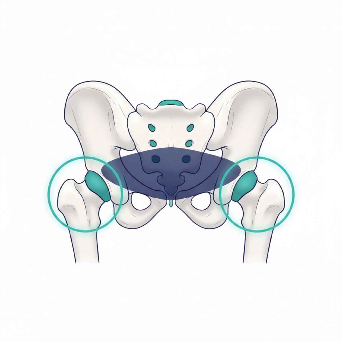
To accurately palpate the gluteus minimus, two primary bony landmarks are essential: the Posterior Superior Iliac Spine (PSIS) and the Greater Trochanter of the femur. The gluteus minimus originates from the external surface of the ilium, between the anterior and inferior gluteal lines, and inserts onto the anterior surface of the greater trochanter. Therefore, it lies deep within the region connecting these two points [5].
Begin by identifying the PSIS, a prominent bony point at the back of the pelvis, and the Greater Trochanter, the large bony prominence on the outer side of the hip. The gluteus minimus resides deep to the gluteus maximus and the tensor fasciae latae (TFL), requiring a careful, deep palpation technique. It is crucial to distinguish it from the gluteus medius, which lies more superficially.
Practice locating these landmarks in a prone position, as this is often the starting point for many therapeutic interventions. Confirming the precise location of these bony guides is the first, non-negotiable step toward successfully isolating the gluteus minimus for effective **myofascial release for low back pain**.
Optimizing Gluteus Minimus Release: Positional Considerations
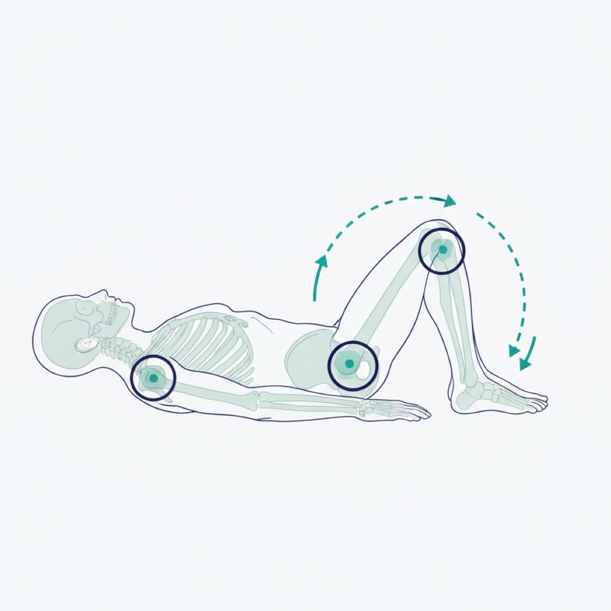
The position of the hip significantly influences the accessibility and tension of the gluteus minimus. One particularly useful position for both assessment and treatment is the 'frog leg' position, which involves hip flexion and external rotation. In this posture, the relative positions of the PSIS and Greater Trochanter shift, altering the orientation and tension of the gluteus minimus [6].
While the 'frog leg' position can make initial palpation more challenging due to the altered anatomical landscape, it can also provide unique access to different parts of the muscle belly or expose areas of increased tension that might be less apparent in a neutral position. Clinicians should practice palpating the gluteus minimus in various hip positions, including flexion, to develop a comprehensive understanding of its presentation and to optimize the effectiveness of **gluteal muscle dysfunction** treatment.
Frequently asked questions
Is fascia release safe for low back pain? — Myofascial release, when performed by a trained clinician, is generally considered safe for addressing low back pain. However, it's essential to consult with a healthcare professional to determine if it's appropriate for your specific condition, especially if you have underlying medical issues.
How long does it take to feel relief from gluteus minimus treatment? — The timeline for feeling relief can vary greatly depending on the individual, the chronicity of the pain, and the specific treatment approach. Some individuals may experience immediate relief, while others might require several sessions to achieve significant improvement. Consistency and adherence to a prescribed treatment plan are key.
Can I perform gluteus minimus release on myself? — While some self-myofascial release techniques exist, accurately targeting a deep muscle like the gluteus minimus requires a thorough understanding of anatomy and skilled palpation, which is best performed by a trained therapist. Attempting to self-treat without proper guidance may lead to ineffective results or potential injury.
References
- [1]Krause F, Wilke J, Vogt L, Banzer W. Intermuscular force transmission along myofascial chains: a systematic review. J Anat. 2016;228(6):910–918.
- [2]Stecco C, Stern R, Porzionato A, et al. Hyaluronan within fascia in the etiology of myofascial pain. Surg Radiol Anat. 2011;33(10):891–896.
- [3]Ajimsha MS, Al-Mudahka NR, Al-Madzhar JA. Effectiveness of myofascial release: a systematic review of randomized controlled trials. J Bodyw Mov Ther. 2015;19(1):102–112.
- [4]Tozzi P, Bongiorno D, Vitturini C. Fascial release effects on patients with non-specific cervical or lumbar pain. J Bodyw Mov Ther. 2011;15(4):405–416.
- [5]Zügel M, Maganaris CN, Wilke J, et al. Fascial tissue research in sports medicine: from molecules to tissue adaptation, injury and diagnostics: consensus statement. Br J Sports Med. 2018;52(23):1497.
- [6]Klingler W, Velders M, Hoppe K, Pedro M, Schleip R. Clinical relevance of fascial tissue and dysfunctions. Curr Pain Headache Rep. 2014;18(8):439.
Related videos
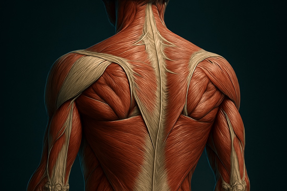
Palpation X-Ray #84 | Is the Sacrum Really a Flat Plate? It's a Wedge, Wide at the Top and Narrowing Downward — Trace Down from the Lumbar Spine and Confirm at Gluteus Maximus
0:56
Dramatic Improvement in One Session: Manual Approach to the Pelvic Girdle, with Palpation of Anatomical Landmarks and Movement Assessment Before and After (Hands-On Demo)
5:48