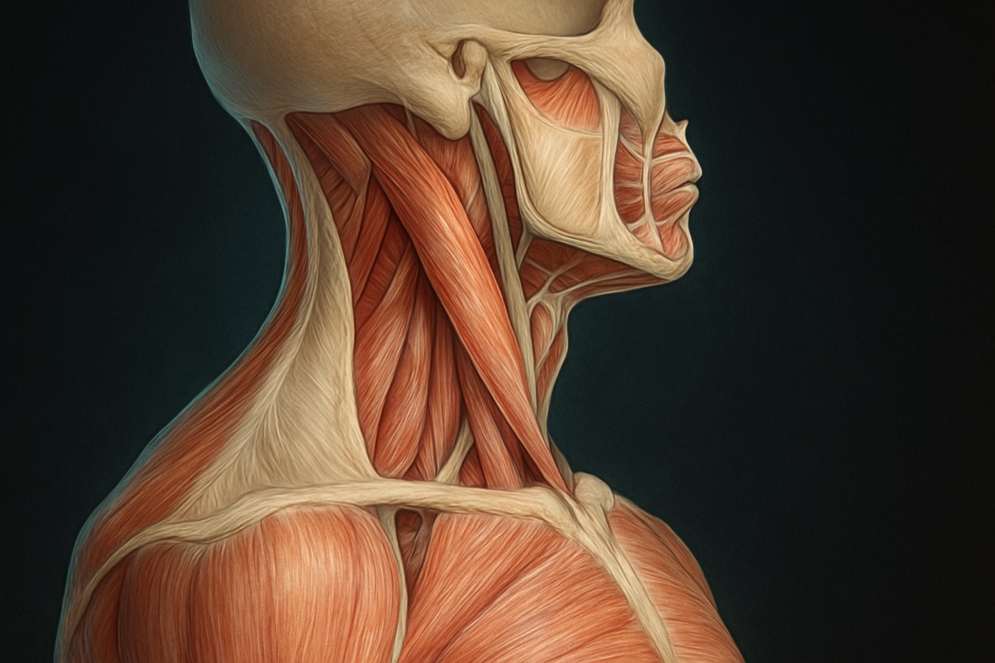
Can 10 Years of Pain Vanish Instantly? Don't Miss Shogo Fujii's Healing Hands
Original title (JP): 10年来の痛みも一瞬で消える?藤井翔悟の神の手を見逃すな!

Supervised by
Shogo Fujii
Physiotherapist (PT) · Founder of the SHOGO FUJII® FASCIA METHOD
View profile →Clinical Article · 6 min read
Anyone who has rolled an ankle and never fully recovered has probably searched for fascia release for ankle sprain pain at some point. In this session, physical therapist Shogo Fujii works with a patient whose inversion ankle sprains started in his school years and never resolved through rest alone. Instead of treating the joint in isolation, Fujii assesses ligament laxity, checks for fracture red flags, and then releases restricted fascia around the peroneal muscles and medial malleolus. The approach illustrates why a joint can look structurally fine on exam yet still hurt with every step.
Why do old ankle sprains keep hurting?
A classic inversion sprain happens when the foot rolls inward, straining the lateral ligaments and the fascia surrounding the peroneal tendons that run along the outside of the ankle. When someone reports repeated sprains since childhood or adolescence, the tissue around that joint is often left lax and less able to signal the ankle's true position in space, a factor linked to chronic ankle instability [1][2].
In the video, Fujii first checks passive laxity by moving the uninjured ankle into an inverted position and noting how far it travels before reaching its endfeel, the natural stopping point of a joint's range. A markedly loose endfeel on the healthy side suggested this patient was simply prone to spraining, which reframed the current complaint as one episode in a longer pattern rather than an isolated injury.
How was the injury assessed before any hands-on work?
Before touching the fascia, Fujii ruled out signs of a more serious injury. He noted the patient could still bear weight and walk on the ankle immediately after the sprain, and that swelling had been minimal, both of which pointed away from a significant ligament tear or fracture. He specifically checked for bony tenderness because an occult fracture can mimic a soft-tissue sprain, and mentioned that persistent swelling or suspected bone injury calls for imaging rather than manual therapy alone.
With structural damage less likely, the remaining pain was traced to two everyday movements: walking and the initial phase of squatting. Reproducing those positions let Fujii see exactly which motion still provoked pain, which he then used as a live test throughout the session rather than relying on the patient's description alone.
How does fascia release target the peroneal fascia?
Fujii located the tender band near the origin of the peroneus longus, close to the fibular head, and applied sustained pressure there while asking the patient to move the ankle into the position that normally caused pain. When the same movement no longer hurt with pressure applied, he treated that spot as the true source of the restriction rather than the ankle joint itself, a diagnostic technique sometimes called provocation-and-release testing [3].
He then worked more locally around the medial malleolus, feeling for a mobile band of tissue and gently working it back into a more normal glide. Myofascial and soft-tissue techniques like this are thought to reduce local tissue density and improve the sliding between fascial layers, which can lessen pain and restore a fuller range of motion in patients with lingering post-sprain symptoms [4][5].
A useful clinical sign Fujii highlights is tissue that visibly softens, or 'gives,' under the same pressure that was painful minutes earlier — he uses this softening, rather than the patient's verbal report alone, as a marker that the restriction has released.
What can patients expect afterward, and are there limits?
Immediately after the release, the patient repeated the walking and foot movements that had been painful and reported the pain was gone. Fujii frames this kind of quick change as a working sign that the fascia, not the joint structure, was driving the symptom, though he's clear this is a same-session finding rather than a guaranteed permanent fix, and encourages using the affected ankle through both inward- and outward-rolling movements afterward to see how it holds up under normal loading [6].
The video also shows the limits of manual technique alone: with an older patient managing a different kind of injury, mobility work had to be adapted to a much slower pace, using support like parallel bars and modified positions rather than the brisk hands-on release used for the ankle sprain. This distinction matters for readers — fascia work is one tool among several, and technique, tissue age, and injury history all shape what a realistic outcome looks like.
Frequently asked questions
Is fascia release safe for an old ankle sprain? — For most people without a suspected fracture, ongoing swelling, or acute instability, targeted fascia work on the peroneal region is considered low-risk, but a clinician should first rule out bony injury or ligament rupture before any manual technique is applied.
Why does an ankle sprain from years ago still hurt? — Fascia and connective tissue around the joint can remain restricted or the surrounding ligaments can stay lax long after the original injury has healed, which keeps certain movements, like the start of a squat or a rolling step, uncomfortable even though imaging may look unremarkable.
Can fascia release replace physical therapy or medical evaluation? — No; it is best used alongside, not instead of, a proper evaluation, and anyone with persistent swelling, instability, or suspected fracture should see a qualified clinician before pursuing manual fascia techniques.
References
- [1]Wilke J, Krause F, Vogt L, Banzer W. What Is Evidence-Based About Myofascial Chains? A Systematic Review. Arch Phys Med Rehabil. 2016;97(3):454–461.
- [2]Krause F, Wilke J, Vogt L, Banzer W. Intermuscular force transmission along myofascial chains: a systematic review. J Anat. 2016;228(6):910–918.
- [3]Stecco C, Stern R, Porzionato A, et al. Hyaluronan within fascia in the etiology of myofascial pain. Surg Radiol Anat. 2011;33(10):891–896.
- [4]Ajimsha MS, Al-Mudahka NR, Al-Madzhar JA. Effectiveness of myofascial release: a systematic review of randomized controlled trials. J Bodyw Mov Ther. 2015;19(1):102–112.
- [5]Tozzi P, Bongiorno D, Vitturini C. Fascial release effects on patients with non-specific cervical or lumbar pain. J Bodyw Mov Ther. 2011;15(4):405–416.
- [6]Zügel M, Maganaris CN, Wilke J, et al. Fascial tissue research in sports medicine: from molecules to tissue adaptation, injury and diagnostics: consensus statement. Br J Sports Med. 2018;52(23):1497.
Related videos
Palpation X-Ray #84 | Is the Sacrum Really a Flat Plate? It's a Wedge, Wide at the Top and Narrowing Downward — Trace Down from the Lumbar Spine and Confirm at Gluteus Maximus
0:56
Dramatic Improvement in One Session: Manual Approach to the Pelvic Girdle, with Palpation of Anatomical Landmarks and Movement Assessment Before and After (Hands-On Demo)
5:48